- Clinical Study
- Identification of Novel Genetic Variants Related to Trabecular Bone Score in Community-Dwelling Older Adults
-
Sung Hye Kong, Ji Won Yoon, Jung Hee Kim, JooYong Park, Jiyeob Choi, Ji Hyun Lee, A Ram Hong, Nam H. Cho, Chan Soo Shin
-
Endocrinol Metab. 2020;35(4):801-810. Published online November 24, 2020
-
DOI: https://doi.org/10.3803/EnM.2020.735
-
-
 Abstract Abstract
 PDF PDF Supplementary Material Supplementary Material PubReader PubReader  ePub ePub
- Background
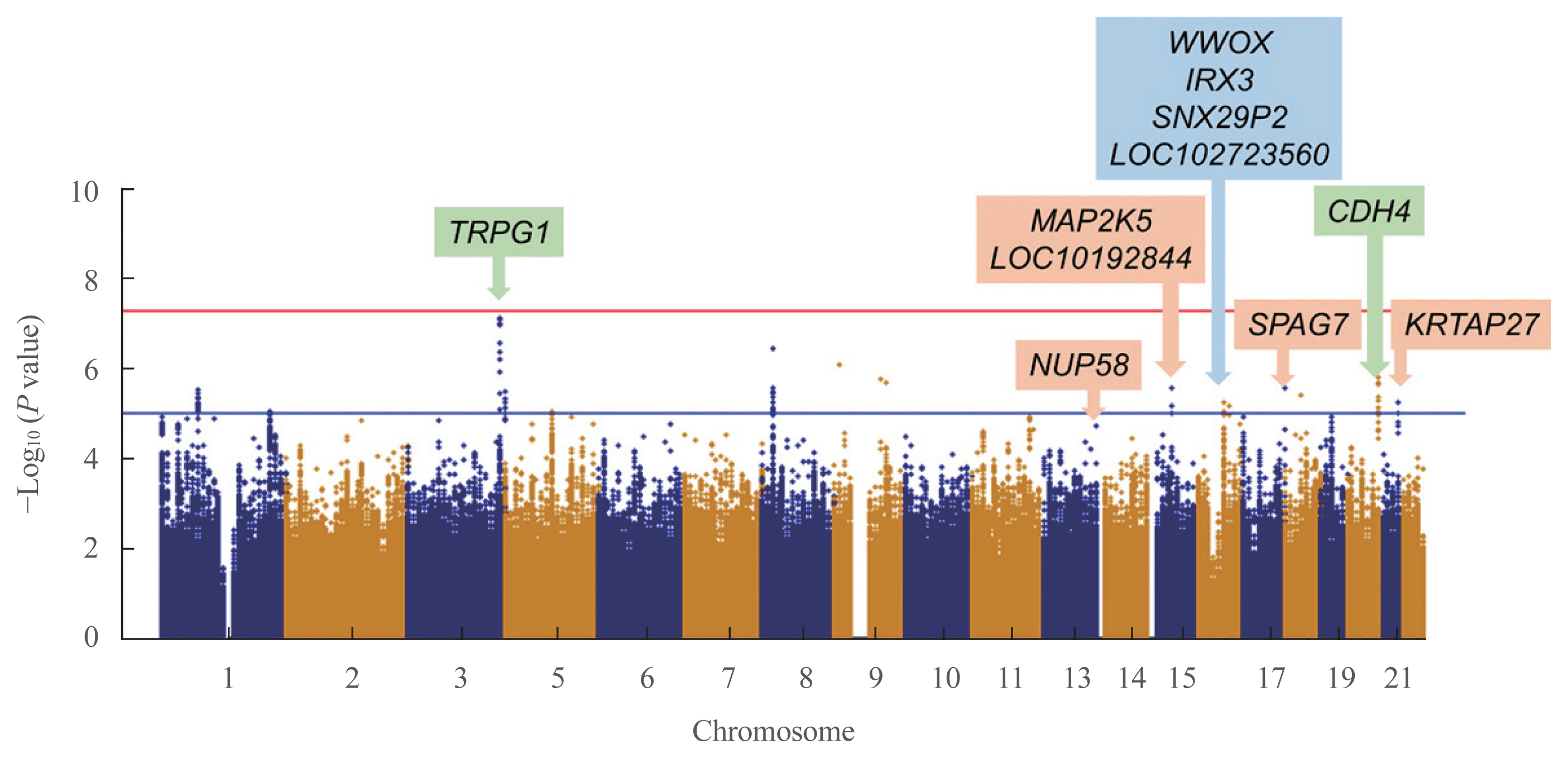
As the genetic variants of trabecular bone microarchitecture are not well-understood, we performed a genome-wide association study to identify genetic determinants of bone microarchitecture analyzed by trabecular bone score (TBS).
Methods
TBS-associated genes were discovered in the Ansung cohort (discovery cohort), a community-based rural cohort in Korea, and then validated in the Gene-Environment Interaction and Phenotype (GENIE) cohort (validation cohort), consisting of subjects who underwent health check-up programs. In the discovery cohort, 2,451 participants were investigated for 1.42 million genotyped and imputed markers.
Results
In the validation cohort, identified as significant variants were evaluated in 2,733 participants. An intronic variant in iroquois homeobox 3 (IRX3), rs1815994, was significantly associated with TBS in men (P=3.74E-05 in the discovery cohort, P=0.027 in the validation cohort). Another intronic variant in mitogen-activated protein kinase kinase 5 (MAP2K5), rs11630730, was significantly associated with TBS in women (P=3.05E-09 in the discovery cohort, P=0.041 in the validation cohort). Men with the rs1815994 variant and women with the rs11630730 variant had lower TBS and lumbar spine bone mineral density. The detrimental effects of the rs1815994 variant in men and rs11630730 variant in women were also identified in association analysis (β=–0.0281, β=–0.0465, respectively).
Conclusion
In this study, the rs1815994 near IRX3 in men and rs11630730 near MAP2K5 in women were associated with deterioration of the bone microarchitecture. It is the first study to determine the association of genetic variants with TBS. Further studies are needed to confirm our findings and identify additional variants contributing to the trabecular bone microarchitecture.
- Clinical Study
- Low Predictive Value of FRAX Adjusted by Trabecular Bone Score for Osteoporotic Fractures in Korean Women: A Community-Based Cohort Study
-
Hana Kim, Jung Hee Kim, Min Joo Kim, A Ram Hong, HyungJin Choi, EuJeong Ku, Ji Hyun Lee, Chan Soo Shin, Nam H. Cho
-
Endocrinol Metab. 2020;35(2):359-366. Published online June 24, 2020
-
DOI: https://doi.org/10.3803/EnM.2020.35.2.359
-
-
5,944
View
-
132
Download
-
9
Web of Science
-
10
Crossref
-
Abstract
PDFPubReader ePub
- Background
The value of the Fracture Risk Assessment Tool (FRAX) and the trabecular bone score (TBS) for assessing osteoporotic fracture risk has not been fully elucidated in Koreans. We conducted this study to clarify the predictive value of FRAX adjusted by TBS for osteoporotic fractures in Korean women.
Methods
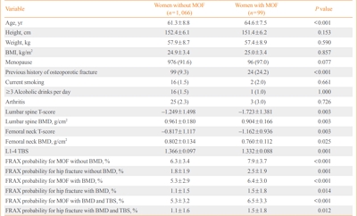
After screening 7,192 eligible subjects from the Ansung cohort, 1,165 women aged 45 to 76 years with available bone mineral density (BMD) and TBS data were enrolled in this study. We assessed their clinical risk factors for osteoporotic fractures and evaluated the predictive value of FRAX with or without BMD and TBS.
Results
During the mean follow-up period of 7.5 years, 99 (8.5%) women suffered major osteoporotic fractures (MOFs) and 28 (2.4%) experienced hip fractures. FRAX without BMD, BMD-adjusted FRAX, and TBS-adjusted FRAX were significantly associated with the risk of MOFs (hazard ratio [HR] per percent increase, 1.08; 95% confidence interval [CI], 1.03 to 1.14; HR, 1.09; 95% CI, 1.03 to 1.15; and HR, 1.07; 95% CI, 1.02 to 1.13, respectively). However, BMD-adjusted FRAX and TBS-adjusted FRAX did not predict MOFs better than FRAX without BMD based on the Harrell’s C statistic. FRAX probabilities showed limited value for predicting hip fractures. The cut-off values of FRAX without BMD, FRAX with BMD, and FRAX with BMD adjusted by TBS for predicting MOFs were 7.2%, 5.0%, and 6.7%, respectively.
Conclusion
FRAX with BMD and TBS adjustment did not show better predictive value for osteoporotic fractures in this study than FRAX without adjustment. Moreover, the cut-off values of FRAX probabilities for treatment might be lower in Korean women than in other countries.
-
Citations
Citations to this article as recorded by  - Update on the utility of trabecular bone score (TBS) in clinical practice for the management of osteoporosis: a systematic review by the Egyptian Academy of Bone and Muscle Health
Yasser El Miedany, Walaa Elwakil, Mohammed Hassan Abu-Zaid, Safaa Mahran
Egyptian Rheumatology and Rehabilitation.2024;[Epub] CrossRef - Comparison of predictive value of FRAX, trabecular bone score, and bone mineral density for vertebral fractures in systemic sclerosis: A cross-sectional study
Kyung-Ann Lee, Hyun-Joo Kim, Hyun-Sook Kim
Medicine.2023; 102(2): e32580. CrossRef - Screening for the primary prevention of fragility fractures among adults aged 40 years and older in primary care: systematic reviews of the effects and acceptability of screening and treatment, and the accuracy of risk prediction tools
Michelle Gates, Jennifer Pillay, Megan Nuspl, Aireen Wingert, Ben Vandermeer, Lisa Hartling
Systematic Reviews.2023;[Epub] CrossRef - Chronic airway disease as a major risk factor for fractures in osteopenic women: Nationwide cohort study
Sung Hye Kong, Ae Jeong Jo, Chan Mi Park, Kyun Ik Park, Ji Eun Yun, Jung Hee Kim
Frontiers in Endocrinology.2023;[Epub] CrossRef - Update on the clinical use of trabecular bone score (TBS) in the management of osteoporosis: results of an expert group meeting organized by the European Society for Clinical and Economic Aspects of Osteoporosis, Osteoarthritis and Musculoskeletal Disease
Enisa Shevroja, Jean-Yves Reginster, Olivier Lamy, Nasser Al-Daghri, Manju Chandran, Anne-Laurence Demoux-Baiada, Lynn Kohlmeier, Marie-Paule Lecart, Daniel Messina, Bruno Muzzi Camargos, Juraj Payer, Sansin Tuzun, Nicola Veronese, Cyrus Cooper, Eugene V.
Osteoporosis International.2023; 34(9): 1501. CrossRef - Comparison of HU histogram analysis and BMD for proximal femoral fragility fracture assessment: a retrospective single-center case–control study
Sun-Young Park, Hong Il Ha, Injae Lee, Hyun Kyung Lim
European Radiology.2022; 32(3): 1448. CrossRef - Association of Trabecular Bone Score-Adjusted Fracture Risk Assessment Tool with Coronary Artery Calcification in Women
Tzyy-Ling Chuang, Yuh-Feng Wang, Malcolm Koo, Mei-Hua Chuang
Diagnostics.2022; 12(1): 178. CrossRef - Risk of osteoporotic fracture in women using the FRAX tool with and without bone mineral density score in patients followed at a tertiary outpatient clinic ‒ An observational study
Maria Helena Sampaio Favarato, Maria Flora de Almeida, Arnaldo Lichtenstein, Milton de Arruda Martins, Mario Ferreira Junior
Clinics.2022; 77: 100015. CrossRef - Comparison of Trabecular Bone Score–Adjusted Fracture Risk Assessment (TBS-FRAX) and FRAX Tools for Identification of High Fracture Risk among Taiwanese Adults Aged 50 to 90 Years with or without Prediabetes and Diabetes
Tzyy-Ling Chuang, Mei-Hua Chuang, Yuh-Feng Wang, Malcolm Koo
Medicina.2022; 58(12): 1766. CrossRef - Application of the Trabecular Bone Score in Clinical Practice
Sung Hye Kong, Namki Hong, Jin-Woo Kim, Deog Yoon Kim, Jung Hee Kim
Journal of Bone Metabolism.2021; 28(2): 101. CrossRef
- Clinical Study
- Radiographic Characteristics of Adrenal Masses in Oncologic Patients
-
Ji Hyun Lee, Eun Ky Kim, A Ram Hong, Eun Roh, Jae Hyun Bae, Jung Hee Kim, Chan Soo Shin, Seong Yeon Kim, Sang Wan Kim
-
Endocrinol Metab. 2016;31(1):147-152. Published online March 16, 2016
-
DOI: https://doi.org/10.3803/EnM.2016.31.1.147
-
-
4,423
View
-
36
Download
-
9
Web of Science
-
8
Crossref
-
Abstract
PDFPubReader
- Background
We aimed to assess the usefulness of pre-contrast Hounsfield unit (HU) and mass size on computed tomography to differentiate adrenal mass found incidentally in oncologic patients. MethodsFrom 2000 to 2012, 131 oncologic patients with adrenal incidentaloma were reviewed retrospectively. Receiver operating characteristic (ROC) curves were applied to determine the optimal cut-off value of the mean HU and size for detecting adrenal metastasis. ResultsThe median age was 18 years, and 80 patients were male. The initial mass size was 18 mm, and 71 (54.2%) of these were on the left side. A bilateral adrenal mass was found in 11 patients (8.4%). Biochemically functional masses were observed in 9.2% of patients. Thirty-six out of 119 patients with nonfunctional masses underwent adrenalectomy, which revealed metastasis in 13. The primary cancers were lung cancer (n=4), renal cell carcinoma (n=2), lymphoma (n=2), hepatocellular carcinoma (n=2), breast cancer (n=1), and others (n=2). The area under the curve for the size and HU for clinically suspicious metastasis were 0.839 (95% confidence interval [CI], 0.761 to 0.900; P<0.001) and 0.959 (95% CI, 0.898 to 0.988; P<0.001), respectively. The cut-off value to distinguish between metastasis and benign masses were 22 mm for size and 20 for HU. ConclusionROC curve results suggest that pre-contrast HU >20 can be used as a diagnostic reference to suggest metastasis in oncologic patients with adrenal masses.
-
Citations
Citations to this article as recorded by - Risk of malignancy in adrenal tumors in patients with a history of cancer
Radosław Samsel, Karolina Nowak, Lucyna Papierska, Edyta Karpeta, Katarzyna Roszkowska-Purska, Wacław Smiertka, Tomasz Ostrowski, Eryk Chrapowicki, Alan Grabowski, Dorota Leszczyńska, Andrzej Cichocki
Frontiers in Oncology.2023;[Epub] CrossRef - Adrenal Tumors Found During Staging and Surveillance for Colorectal Cancer: Benign Incidentalomas or Metastatic Disease?
Mio Yanagisawa, Dania G. Malik, Thomas W. Loehfelm, Ghaneh Fananapazir, Michael T. Corwin, Michael J. Campbell
World Journal of Surgery.2020; 44(7): 2282. CrossRef - Predictive factors for adrenal metastasis in extra‐adrenal malignancy patients with solitary adrenal mass
Kyeong‐Hyeon Byeon, Yun‐Sok Ha, Seock‐Hwan Choi, Bum Soo Kim, Hyun Tae Kim, Eun Sang Yoo, Tae Gyun Kwon, Jun Nyung Lee, Tae‐Hwan Kim
Journal of Surgical Oncology.2018; 118(8): 1271. CrossRef - Combining Washout and Noncontrast Data From Adrenal Protocol CT
Chaan S. Ng, Emre Altinmakas, Wei Wei, Payel Ghosh, Xiao Li, Elizabeth G. Grubbs, Nancy A. Perrier, Victor G. Prieto, Jeffrey E. Lee, Brian P. Hobbs
Academic Radiology.2018; 25(7): 861. CrossRef - Evaluation of quantitative parameters for distinguishing pheochromocytoma from other adrenal tumors
Youichi Ohno, Masakatsu Sone, Daisuke Taura, Toshinari Yamasaki, Katsutoshi Kojima, Kyoko Honda-Kohmo, Yorihide Fukuda, Koji Matsuo, Toshihito Fujii, Akihiro Yasoda, Osamu Ogawa, Nobuya Inagaki
Hypertension Research.2018; 41(3): 165. CrossRef - Articles inEndocrinology and Metabolismin 2016
Won-Young Lee
Endocrinology and Metabolism.2017; 32(1): 62. CrossRef - The Diverse Clinical Presentations of Adrenal Lymphoma
Awais Masood, Anna Tumyan, Daniel R. Nussenzveig, Dara N. Wakefield, Diana Barb, Hans K. Ghayee, Naim M. Maalouf
AACE Clinical Case Reports.2017; 3(4): 307. CrossRef - Optimal follow-up strategies for adrenal incidentalomas: reappraisal of the 2016 ESE-ENSAT guidelines in real clinical practice
A Ram Hong, Jung Hee Kim, Kyeong Seon Park, Kyong Young Kim, Ji Hyun Lee, Sung Hye Kong, Seo Young Lee, Chan Soo Shin, Sang Wan Kim, Seong Yeon Kim
European Journal of Endocrinology.2017; 177(6): 475. CrossRef
- Free T4 is Negatively Correlated with Non-alcoholic Fatty Liver Disease in Euthyroid Women.
-
Eon Ju Jun, Hyun Sook Kim, Hyue Kyung Chung, Ji Hyun Lee, Sae Rom Kim, Eui Dal Jung
-
J Korean Endocr Soc. 2009;24(2):87-92. Published online June 1, 2009
-
DOI: https://doi.org/10.3803/jkes.2009.24.2.87
-
-
Abstract
PDF
- BACKGROUND
Thyroid hormones play an important role in the regulation of lipid and carbohydrate metabolism and the body mass index (BMI), which all affect non-alcoholic fatty liver disease (NAFLD). In a previous study, we demonstrated that free T4 was negatively associated with the BMI in euthyroid women. However, there is still uncertain as to whether the thyroid function within the normal range is associated with NAFLD and liver function abnormalities. We sought to evaluate the thyroid function (free T4, TSH) and its possible relationship with NAFLD in euthyroid women. METHODS: A total of 835 euthyroid, non heavy alcoholics women who visited the Daegu Catholic University University Medical Centre for primary health screening from January 1, 2006 to December 31, 2006 participated in this cross-sectional study. The women who were not euthyroid or heavy alcoholics (> 70 g/week in women according to the DSM-IV), there was no known history of diabetes mellitus, the fasting blood glucose was more than 5.55 mmol/L and those who had viral hepatitis were excluded. Hepatic ultrasonography scanning was performed in all the participants by a single experienced radiologist. The TSH, free T4, BP, fasting glucose, serum liver enzymes (AST, ALT, GGT, T-bilirubin), lipid profiles [total-cholesterol, triglyceride (TG), HDL-C, LDL-C] and NAFLD were evaluated. RESULTS: Euthyroid women with NAFLD had lower free T4 levels than did the euthyroid women without NAFLD. After adjustment for age and BMI, free T4 was negatively correlated with TG, but free T4 was positively correlated with the total serum bilirubin. Free T4 was not correlated with the serum AST, ALT and GGT. After adjustment for age, the BMI, the fasting glucose, the GGT and free T4, but not TSH, were significantly negatively correlated with NAFLD. CONCLUSION: We demonstrated a negative correlation between free T4 and NAFLD in euthyroid women. This finding suggests lower levels of free T4 is associated with NAFLD in euthyroid subjects.
- A Case of Idiopathic Central Diabetes Insipidus together with Primary Empty Sella and Combined Pituitary Hormone Deficiency.
-
Sun Young Ahn, Kyu Hwan Bae, Myung Hwan Kim, Ji Hyun Lee, Ho Sang Shon, Eui Dal Jung
-
J Korean Endocr Soc. 2007;22(4):272-276. Published online August 1, 2007
-
DOI: https://doi.org/10.3803/jkes.2007.22.4.272
-
-
Abstract
PDF
- Central diabetes insipidus is a heterogeneous condition that is characterized by polyuria and polydipsia, and this is due to a deficiency of arginine vasopressin. Central diabetes insipidus is rare in children and young adults, and up to 50 percent of cases are idiopathic. Genetic abnormalities in the homeobox genes have recently been shown, on sellar magnetic resonance imaging, to be associated with combined pituitary hormone deficiency with pituitary defect. We report here on a 44-year-old female who suffered from polydipsia, polyuria and primary amenorrhea since childhood. She was diagnosed with idiopathic central diabetes insipidus together with primary empty sella and combined pituitary hormone deficiency. On the genetic analysis, she was proven to have a point mutation of the PROP-1 gene, which is known as a cause of combined pituitary hormone deficiency.
- A Case of Primary Aldosteronism with Aortic Dissection.
-
Jung Hyun Seo, Ji Young Kim, Young Sup Kim, Wan Suk Kim, Jin Hyang Shin, Dong Jik Ahn, Yoon Young Cho, Sung Hwan Park, Jae Bok Park, Hyun Dae Yoon, Ji Hyun Lee, Ho Sang Shon
-
J Korean Endocr Soc. 2004;19(4):452-457. Published online August 1, 2004
-
-
-
Abstract
PDF
- Hypertension and atherosclerosis are the most important factors contributing to the development of aortic dissection. Primary aldosteronism is a rare cause of hypertension. The concurrence of aortic dissection is very rare in primary aldosteronism. However, when aortic dissection is found as a life-threatening complication of primary aldosteronism, then the diagnosis of primary aldosteronism is important because the therapeutic intervention can be curative. Only 3 cases of primary aldosteronism with aortic dissection have been reported in the literature. We report here on a case of primary aldosteronism with aortic dissection, which was treated by laparoscopic adrenalectomy. We lowered the blood pressure with antihypertensive drugs and potassium replacement was done to treat the aortic dissection. After stabilization of aortic dissection, we removed his left adrenal mass by laparoscopic adrenalectomy. Postoperatively, the patient's blood pressure has been within normal limits and the serum potassium increased to a normal level without supplementation. The aortic dissection has remained in a stable state
- Association of Polymorphism in beta3-Adrenergic Receptor Gene with Fat Distribution.
-
Tae Sung Yun, Yong Deuk Kim, Hye Soon Kim, Mi Jung Kim, Young Sung Suh, Jung Hyeok Kwon, Jin Soo Choi, Jung Guk Kim, Sung Woo Ha, Bo Wan Kim, Kyu Chang Won, Hyong Woo Lee, Ho Sang Shon, Ji Hyun Lee, Hyun Dae Yoon, Won Ho Kim, Young Gil Yun, In Kyu Lee
-
J Korean Endocr Soc. 2003;18(2):184-192. Published online April 1, 2003
-
-
-
Abstract
PDF
- BACKGROUND
Reasons for obesity include environmental factors and, more largely so, genetic factors. There have been many studies on these genetic factors. So far, genes related to obesity such as Leptin, Uncoupling Protein(UCP), Peroxisome proliferator activated receptor-gamma(PPAR-gamma), and Beta3-adrener-gic receptor(beta3-AR) gene have been discovered. Among these, beta3-AR is expressed in visceral adipose tissue and is thought to contribute to the regulation of resting metabolic rate and lipolysis. The missense mutation of beta3-AR gene, resulting in replacement of tryptophan by arginine at position 64(Trp64Arg), is associated with decreased resting metabolic rate and weightgain. We performed this study to determine if Trp64Arg polymorphism of beta3-AR gene is associatedwith obesity in Koreans. METHOD: We investigated the relationship between the beta3-AR gene mutation and body mass index (BMI), waist circumference, hip circumference, waist to hip ratio(WHR), area of subcutaneous fat, area of visceral fat, visceral to subcutaneous fat ratio(VSR), and lipid profile. 198 subjects were included in this study of which 97 were of normal weight and 101 were obese. Anthropometric data was obtained from physical examination and medical records. RESULT: In the cases of beta3-AR gene mutation of the obese group, the ratio of Trp/Arg and Arg/Arg are 43% and 5%, respectively, which were higher than the normal group(36%, 1%), although a statistical significant was not found. There was significant difference in the are of subcutaneous fat. Normal group(Trp/Trp) measured at 213.9+/-109.6cm2 versus 244.0+/-127.7cm2 (Trp/Arg) and 323.9+/-189.9cm2(Arg/Arg) for the mutation groups. Circumference of waist, circumference of hip, WHR, area of visceral fat, and VSR were higher in the mutation groups than in normal subject, but not significantly different. CONCLUSION: These results suggest that a genetic mutation in the beta3-AR gene can affect body fat composition, and is associated with obesity in Korean adults.
- A Case of Graves' Disease Associated with Guillain-Barre Syndrome.
-
Ji Hyun Lee, Ki Sung Ahn, Sang Chae Lee, Jung Dong Bae, Yong Bum Park, Soo Mi Keum, Jin Hyung Park, Jong Won Choi, Ji Yong Choi, Sung Kook Jang, Ho Sang Son
-
J Korean Endocr Soc. 1997;12(4):614-620. Published online January 1, 2001
-
-
-
Abstract
PDF
- Graves disease, an autoimmune endocrine disorder, which causes defects in cellular and humoral immunity, is associated with insulin-dependent diabetes mellitus, Addisons disease, pemicious anemia, and rheumatoid arthritis. Graves disease is associated with various neuro-muscular disorders, such as myopathy, exophalmous oculopathy, periodic paralysis, myastenia gravis and rarely Guillain-Barre syndrome. Guillain-Barre syndrome is considered as an autoimmune disease which can occur concurrently with other autoimmune disorders. This syndrome is characterized by segmental demyelination and axonal degeneration in electrophysiology due to autoantibody to nervous systems via cellular and humoral autoimmunity. In Graves disease, the exact mechanism of the associated Guillain-Barre syndrome is not well understood but it is considered that the autoimmunity is the leading cause of development of both diseases. A 37 year-old man had suffered from thyrotoxic symptoms and progressive symmetrical muscular paralysis. In nerve conduction velocity studies, the result shows peripheral neuropathy; axonopathy; myelinopathy; motor nerve and sensory nerve derangement; right first sacral nerve neuropathy; and decreased CMAP amplitude. The patient was treated with propylthiouracil and high dose intravenous immunoglobulin (400mg/kg/day for Sdays). He responded to the therapy well and became euthyroid state with improvement of muscle weakness. We report a case of Graves' disease associated with Guillain-Barre syndrome with brief review of literature which shows a possible relationship between both diseases.
- A Study About Correlation Between Urinary Androgen Metabolites and Bone Mineral Density in Psstmenopausal Women.
-
Kyoung Rae Kim, Ji Hyun Lee, Sung Kil Lim, Young Jun Won, Seok Ho Kwon, Bong Soo Cha, Young Duk Song, Hyun Chul Lee, Kap Bum Huh, Su Youn Nam, Bong Chul Jung
-
J Korean Endocr Soc. 1997;12(3):450-461. Published online January 1, 2001
-
-
-
Abstract
PDF
- BACKGROUND
Positive correlations between bone mass and androgen levels have been observed in premenopausal and postmenopausal women as well as in men. Androgen production was decreased in women with osteoporosis compared to that in age-matched controls. We hypothesized that androgen metabolism might be also deranged in osteoporosis. To clarify our hypothesis, we investigated the relationship between urinary metabolites of androgen and bone mineral density (BMD) in Korean postmenopausal osteoporotics. METHODS: We examined the anthropometry and bone turnover marker in 67 postmenopausal women. BMD was measured by dual energy X-ray absorptiometry (DEXA). Serurn levels of estrone, estradiol, free testosterone were measured by radioirnmunoassay and serum level of sex hormone binding globulin (SHBG) was measured by two site immunoradiometric assay. The urinary metabolites of androgen were determined by gas chromatography-mass spectrometry (GC-MS) at Korean Institute of Science and Technology Doping Control Center. RESULTS: 1. Spinal BMD had a positive correlation with height (r 0.3049, p<0.05), weight (r=0.4114, p<0.001) and body mass index (BMI, r=0.2638, p<0,05). 2. Spinal and femoral neck BMD had no correlation with serum levels of estrone, estradiol and ten major urinary metabolites of androgen, but serum free testosterone had positive correlation with spinal BMD (r=0.3622, p<0.01) and SHBG had negative correlation with femoral neck BMD (r=-0.2625, p< (0.05). 3. Serum free testosterone in osteoporotics was lower than non-osteoporotics with spinal BMD (p<0.05) and SHBG in patients with osteopenia was higher than non-osteopenic subjects with femoral neck BMD (p <0.05). 4. In multiple stepwise regression analysis, weight and serum free testosterone were statistically significant for spinal BMD (R =0.3072). As for femoral neck BMD, weight was the independent determinant (R 0.1307). 5. Serum level of osteo#ealcin and urinary deoxypyridinoline/creatinine had a positive correlation with urinary 11-ketoandrosterone (p<0.05). SHBG was positive correlation with osteocalcin (r=0.3190, p<0.05). 6. Serum free testosterone (r=-0.2740, p<0.05) decreased with aging. CONCLUSION: Our data suggest that androgen metabolism is not deranged in osteoporotics, but serum free testosterone is important than estrogen on postmenopausal osteoporosis after 5-10 years menopause.
- Estrogen Receptor Gene Polymorphism, Urinary Estrogen Metabolites and Bone Mineral Density in Korean Postmenopausal Women.
-
Ji Hyun Lee, Sung Kil Lim, Young Jun Won, Seok Ho Kwon, Bong Soo Cha, Young Duk Song, Hyun Chul Lee, Kap Bum Huh
-
J Korean Endocr Soc. 1996;11(4):468-478. Published online November 7, 2019
-
-
-
Abstract
PDF
- Background
Estrogen status is important for maintaining the homeostasis of bone. Estrogen has direct effects on bone cells, through binding to the high-affinity estrogen receptor. Several recent studies suggest that there might be genetically determined variations in biosynthesis and function of estrogen receptor in postmenopausal osteoporosis. Also the main cause of postmenopausal osteoporosis is decreased level of serum estrogen, whereas there had been some suggestion that the remaining estrogen have some effect on bone metabolism after menopause. We investigated the relationship between estrogen receptor gene PvulI polymorphism and bone mineral density(BMD), and the relationship between 18 urinary metabolites of estrogen and BMD in Korean postmeno- pausal osteoporosis. Methods: We examined the PvuII polymorphism of the estrogen receptor gene in 5' upstream region and the first intron by restrietion frapnent length polymorphism analysis in 62 postmeno- pausal wornen, BMD was measured by DEXA. The urinary estrogen metabolites were determined by GC/MS(Gas Chromatography-Mass Spectrometry) at Korean Institute of Science and Techno- logy Doping Control Center. Results: BMD of the spine and the femoral neck correlated with body weight, height, body mass index as we expected. There was no polymorphism of PvuII restriction site on 5 upstream region of estrogen receptor gene. Whereas the prevalen~ee of the PP, Pp, pp genotype in the first intron of estrogen receptor was 12.9%, 45.2%, 41.9%, respectively. But, there was no correlation between PvuII genotype and the spinel and femoral neck BMD. 2(OH)E2 among 18 urinary metabolites of estrogen, showed a negative correlation with the spinal and femoral neck BMD(r =-0.2551, p<0.05, and r =-0.3341, p<0.01, respectively), and the ratio of 16a(OH)E2/2(OH)E1> revealed a positive correlation with the spinal BMD(r =0.3057, p<0.05). In stepwise multiple regression analysis, body weight, 2(OH)E2, 16a(OH)E1, 2(Meo)E1 were independent predictors of the spinal bone density, and body weight and 2(OH)E2 were independent predictors of the femoral neck bone density. Conclusion: These results suggested that restrietion fragment length polymorphism analysis of the estrogen receptor gene with PvuII restriction enzyme was not helpful for early detection of patients at risk of developing osteoporosis. However, the ratio of 16-hydroxylation to 2-hydroxylation of estrogen metabolism was reduced in postmenopausal women and high catecholestrogen formation might be a greater risk factor for osteoporosis.
- A Case of Giant Adrenal Adenoma Presenting Primary Aldosteronism.
-
Ji Hyun Lee, Bong Soo Cha, Moon Suk Nam, Young Duk Song, Sung Kil Lim, Hyun Chul Lee, Kap Bum Huh, Hyung Chan Suh, Young Hwa Choi, Jae Min Park, Jung Soo Park, Soon Won Hong, Dong Hwan Shin
-
J Korean Endocr Soc. 1996;11(3):348-354. Published online November 7, 2019
-
-
-
Abstract
PDF
- Primary aldosteronism is a syndrome chracterized by hypokalemic alkalosis and hypertension. Small sized adrenal cortical adenomas have been the major cause of this syndrome in most of the patients. However, if the adrenal mass is larger than 6cm in diameter and with irregular consistency, malignancy is more favored. We experienced a patient who had a giant adrenal adenoma with primary aldosteronism. A 24-year-old female presented with hypertension, hypokalemia, low plasma renin, and high plasrna aldosterone levels, was found to have a 6×5.5×5 cm sized left adrenal tumor by MRI. Her clinical laboratory feature did not revealed any evidence of Cushing's syndrome or pheochromocytoma. Preoperatively adrenal carcinoma presenting pure adrenal aldosteronism was suspected due to large size and heterogenous signal character of the adrenal mass in radiologic study. At operation well encapsulated, round giant adrenal tumor weighing 65gm(4.5×4×4 cm) was removed. There was no evidence of metastasis with return of adrenal function to normal after surgery. Benign adrenal adenoma was confirmed by the gross morphology and the histologic features.
- A Case of Adult Fanconi Syndrome with Hypophosphatemic Osteomalacia.
-
Ji Hyun Lee, Young Sup Byun, Bong Soo Cha, Moon Suk Nam, Young Duk Song, Sung Kil Lim, Kyung Rae Kim, Hyun Chul Lee, Kap Bum Huh, Jin Kim, Jong In Yook
-
J Korean Endocr Soc. 1996;11(1):93-101. Published online November 7, 2019
-
-
-
Abstract
PDF
- The Fanconi syndrome is characterized by generalized disturbance of tubular function. It leads to excessive losses of amino acids, glucose, phosphate, bicarbonate, and other organic and inorganic substrates handled by the proximal tubules. The metabolic consequences are acidosis, hypophosphatemia, hypocalemia, dehydration, rickets, osteomalacia, osteoporosis, and growth retardation. This syndrome may either be congenital or acquired, primary or secondary. Acquired Fanconi syndrome may result from multiple myeloma, Wilsons disease, primary amyloidosis, light chain nephropathy, and heavy metal poisoning such as lead, mercury, and cadmium. A 33-year-old female presented with multiple bone pain, and progressive proximal muscle weakness for 15 months. The blood urea nitrogen, creatinine, calcium, phosphate, and uric acid were 12.1 mg/dL, 1.5 mg/dL, 8.4 mg/dL, 1.8 mg/dL, and 1.7 mg/dL, respectively. The urine volume, protein, calcium, phosphate, and creatinine clearance were 2,330 ml, 343.7 mg, 146 mg, 424 mg, and 44.6 ml/min, respectively in 24 hour collection urine study. The tubular reabsorption rate of phosphate was decreased. In arterial blood gas analysis study, pH was 7.348, bicarbonate was 17.6 mmol/L, which means metabolic acidosis. In chest X-ray, fracture was seen in eighth and ninth left ribs. The whole body bone scan revealed hot uptake at both first and second ribs, right third rib, both eighth and ninth ribs, left sacroiliac joint and right hip joint. Bone densitometry showed moderate osteopenia in spine and femur neck. After NE4Cl loading, the urine pH was decreased below 5.0 at two and third hour, which means proximal renal tubular acidosis. Amino acid such as, hydroxyproline, threonine, serine, asparagine, glutamine excreted much more than normal in 24 hour urine. Bone biopsy showed the presence of increased osteoid volume and osteoid seam width and marked decreased mineral appositional rate as evidence for osteomalacia. The patients symptoms, including bone pain and proximal muscle weakness, were relieved after supplement of calcitonin, Vitamin D and calcium carbonate. We report a case of Fanconi syndrome with hypophosphatemic osteomalacia with brief review of literature.
- Clinical and Endocrinologic Differences between Prolactinoma and Pseudoprolactinoma Proven by Immunohistochemical Study.
-
Jae Wha Jo, Eun Jig Lee, Moon Suk Nam, Su Youn Nam, Young Duk Song, Hyun Chul Lee, Kap Bum Huh, Tae Seung Kim, Sun Ho Kim, Kyung Rae Kim, Bong Soo Cha, Ji Hyun Lee, Sung Kil Lim
-
J Korean Endocr Soc. 1995;10(4):362-369. Published online November 7, 2019
-
-
-
Abstract
PDF
- Hyperprolactinemia is the most common hypothalamo-pituitary disorder encountered in clinical endocrinology. Excluding the drug-induced hyperprolactinemia, the most common cause of this disorder is a pituitary tumor. Prolactinoma is mainly made up of prolactin-secreting cells but pseudoprolactinoma is tumor that does not secrete prolactin itself. The pseudoprolactinoma interrupts the flow of prolactin inhibiting factor, dopamine, from the hypothalamus through the pituitary stalk to the normal pituitary. The differentiation prolactinoma from pseudoprolactinoma is vitally important since true prolactinomas are most commonly responded well in terms of tumor shrinkage to medical treatment using dopamine agonist therapy, whereas pseudoprolactinomas do not. Thus surgical treatment is clearly indicated as first-line treatment if we know that a lesion is a pseudoprolactinoma. We compared prolactinoma with pseudoprolactinoma in clinical and endocrinologic characteristics of 48 cases after immunohistochemical diagnosis. We could not find any differential point of both tumors in clinical and radiological characteristics although some differences were exist. But we had found the relationship between the mean level of pretreatment serum prolactin and the presence of positive immunohistochemical stain for prolactin. The pretreatment serum prolactin level was significantly higher in patients with tumors showing many prolactin immunohistochemical staining cells than in those with none(p<0.05). When the pretreatment serum prolactin exceeded 100ng/ml, the tumors contain 94% of prolactin positive cells in stain. So, if the pretreatment serum prolactin exceeds 100ng/ml, we primarily suspect prolactinoma and medical treatment should be considered. If the pretreatment level below 100ng/ml, we suspect pseudoprolactinoma and surgical treatment should be considered.
- A Case of Idiopathic Juvenile Osteoporosis.
-
Moon Suk Nam, Young Duk Song, Sung Kil Lim, Hyun Chul Lee, Kap Bum Huh, Young Joon Weon, Bong Soo Cha, Ji Hyun Lee, Jung Ho Lee
-
J Korean Endocr Soc. 1994;10(3):278-283. Published online November 6, 2019
-
-
-
Abstract
PDF
- Idiopathic juvenile osteoporosis is a rare disease of heterogenous etiology and occurs on children between the age of 8 and 15. Manifestations include bone pain, fractures in minimal trauma, reduced bone density at areas of new bone growth, and loss of height. It is important to exclude other causes of osteoporosis.We experienced a case of a 14 year old boy with idiopathic juvenile osteoporosis. He had suffered from pain in the back and difficulty on walking for two months. Radiologic finding of the thoracolumbar area of the spine showed generalized severe osteoporosis and multiple vertebral collapse. We could not find the causes of osteoporosis in biochemical study, bone marrow study, skin biopsy and hormonal study. He was treated with alphacalcidol and CaCO_3. After 4 month of initial management, his subjective symtoms were improved and we did not find any signs of progression of disease. On bone mineral density measured after 26 month, we observed markedly increased bone mineral density.We report our experience of follow up of this case and review with the disease reported in the literature.
- Complication and Prognosis of Craniopharyngioma According to the Age of Onset.
-
Eun Jig Lee, Moon Suk Nam, Young Duk Song, Sung Kil Lim, Hyun Chul Lee, Kap Bum Huh, Kyung Rae Kim, Kun Hoon Song, Bong Soo Cha, Ji Hyun Lee
-
J Korean Endocr Soc. 1994;10(3):262-272. Published online November 6, 2019
-
-
-
Abstract
PDF
- Craniopharyngioma is the most common tumor involving the hypothalamo-pituitary area in childhood and adolescence. Recently, we carried out collective review of 70 patients with craniopharyngioma treated from January 1980 to December 1994 in order to inverstigate the endocrine outcome and survival according to the age of onset.The following results were obtained:1) The male to female ratio was 1:1. Age at diagnosis ranged from 2 to 64 years(mean age: 23) with the greatest frequency in the 2nd decade of life(28.6%). Of the 70 cases, the first group, 27 cases were under the age of 15, and the other group, 43 cases were over 15 year-old.2) The most common symptom at diagnosis in both groups was headache. In the adult group, symptoms related to hypogonadism(amenorrhea, decreased libido, galactorrhea etc.) were not uncommon. The lag of time between onset of symptom and hospital visit ranged from 3 days to 156 months(mean: 20 months).3) The main site of tumor was suprasellar region in both groups. The most common CT finding in both groups was calcification in sella turcica.4) In pre-operative combined pituitary function test, the most common, abnormal responses were shown in growth hormone and thyroid stimulating hormone in both groups. In addition, prolactin frequently showed abnormal response in the adult group.In post-operative combined pituitary function test, more hormones tended to reveal abnormal response in the group treated with surgery plus radiation therapy.5) The operation by subtotal removal followed by radiation therapy was the most commonly used method in treatment of both groups. After treatment, panhypopituitarism was occurred more frequently in the group treated with RT after surgery than those treated with surgery alone, but the difference was not statistically significant(p=0.136 in childhood, 0.436 in adults). Except the cases with panhypopituitarism, the most commonly encountered endocrine abnormalities were growth retardation in the children group, and hypogonadism in adult. The recurrence was clinically observed in 11 cases. The recurrence rate were 11.1% in children, and 18.6% in adult respectively. The mean time from the initial treatment to recurrence was 23 months. There was no significant difference in recurrence rate between the group treated with RT after subtotal removal and the group treated with total removal(p=0.475).The overall five-year survival rate after treatment was 82.8%. According to the treatment modalities, the patients undergone RT after subtotal removal survived much longer than those treated with other modalities such as subtotal removal only or total removal, but the differences in survival were not statistically significant(Log rank test, p=0.0539).
|